What is calcific tendinitis?
Rotator cuff calcific tendinitis, as the term suggests, describes the deposition of calcium into the body of a tendon which can then cause an inflammatory reaction that causes pain and dysfunction. Calcium hydroxyapatite is the type of calcium that gets deposited, and these can be quite small and innocuous, however they can grow over time and become painful. If you can imagine toothpaste that has been left out and reacted with the surrounding air – leaving a crusty shell with a softer centre, then you can imagine how it is inside your tendons.
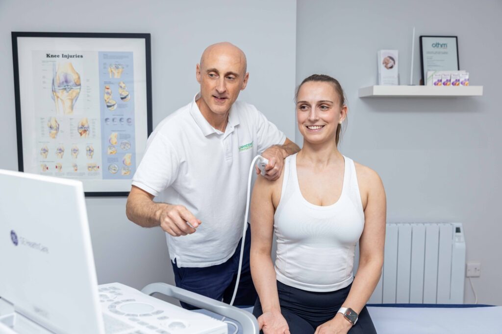
Where does it calcific tendinitis occur?
Calcium deposits can occur in many different tendons. They tend to accumulate in the supraspinatus and or infraspinatus tendons in the shoulder, which can cause pain. They can also accumulate in the patella tendon, Achilles tendon and proximal hamstring tendon. We are not entirely sure why the body deposits calcium in tendons, but it could be to do with a stress reaction in the tendon that leads to calcium deposition. Calcium deposits can be seen very clearly on ultrasound and often they are stable and asymptomatic. For example, they can often be seen in both shoulders, with one shoulder causing pain but no the other. See our shoulder pain clinic for more on how we can help…
What are the symptoms of shoulder calcific tendinitis?
There are two types of rotator cuff calcific tendinitis presentation with regards to shoulder calcific tendinitis 1) a very acute severe presentation or 2) an impingement type presentation.
Severe acute calcific tendinitis
The very acute severe rotator cuff calcific tendinitis patient will often have already been to their GP or even been to A&E as the pain is so bad. They have gross loss of shoulder movement and extreme / severe pain and can’t sleep at night. They are often mistakenly told they have a frozen shoulder. Very little to no testing will be possible in the assessment session at it is too painful. What is happening during this stage is the the calcific deposit is usually undergoing a resorptive phase which means the immune system is breaking down and removing the deposit. This is very well seen on diagnostic ultrasound. These generally require a steroid injection for pain relief.
Impingement calcific tendinitis
The impingement type of rotator cuff calcific tendinitis often feel that they have something blocking a particular movement such as taking a t-shirt off or lifting the arm above the head (abduction or external / internal rotation) and have to click or flick their shoulder to be able to move it. Range of movement and strength are usually good but there will be positive impingement tests. This impingement type of shoulder calcific tendinitis is well seen as a stable calcific deposit in the rotator cuff tendon (usually the supraspinatus and or infraspinatus tendons) on Ultrasound.
What treatments are available for calcific tendinitis?
There are two main types of non-invasive treatment for rotator cuff calcific tendinitis. Shockwave therapy can be useful for the impingement type, where the deposit is stable and mildly irritable, it could also be used for the acute resorptive type. We recommend focused shockwave for rotator cuff calcific tendinitis as this delivers higher energy sound waves to the shoulder and can resolve the problem sooner than radial shockwave.
Ultrasound guided injections for calcific tendinitis
There are different ultrasound guided injections that can be used for calcific tendinitis of the shoulder. For someone who is experiencing an acute bout of severe calcific tendinitis it might be that we start with a steroid injection in the sub-acromial bursa simply for pain relief. This can settle the symptoms down and allow you to get sleep and generally start to use your arm again. It might be in time that body resolves the problem and gets rid of the calcium deposit, however it can sometimes remain a little sore which might benefit from shockwave or a barbotage / lavage process.
For those who have the impingement type of calcific tendinitis it might also be that you have a steroid injection into the bursa to settle any pain, however if shockwave has not helped to remove the calcium you could have a barbotage / lavage procedure. First, we would inject some local anaesthetic into the shoulder bursa to numb the area. Then we would try to either break up the calcium deposit with a needle tip and / or try to use saline to back wash the calcium deposit back into the syringe. Lastly, we would inject a little steroid into the bursa for pain relief.
There is also the option of having the calcium deposit surgically removed and this would involve speaking with a surgeon. For those suffering rotator cuff calcific tendonitis we have a specialist shoulder appointment available at our clinics in Wandsworth Town, Clapham Junction and Battersea Nine Elms and Vauxhall. This includes and assessment, ultrasound scan, strength assessment with VALD force decks and dynamometers before we put together a comprehensive treatment plan including focused or radial shockwave, manual therapy, exercises therapy and ultrasound guided injections where necessary.